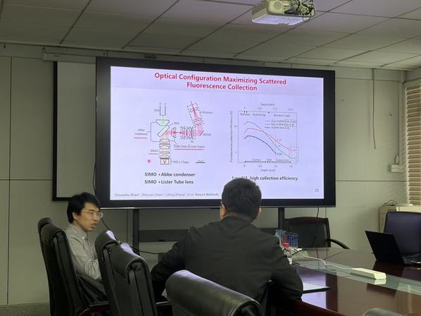
On the morning of April 28, Zhejiang University's College of Biomedical Engineering & Instrument Science (CBEIS) held the 39th Lv Weixue Academic Forum in Room 420 on the fourth floor of the Zhou Yiqing Building. The forum featured an invited academic talk by Dr. Zhao Chunzhu, Researcher at the College of Future Technology, Peking University, and the National Biomedical Imaging Center, Peking University, titled Opening a New Window into Deep-Brain Observation: Innovation and Applications of Miniaturized Multiphoton Imaging Technology. Faculty and graduate students working in related fields attended the forum.
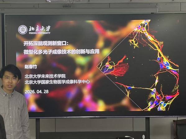
Dr. Zhao Chunzhu has long focused on research in optical brain-computer interfaces and multiphoton microscopy imaging. Centered on the important scientific challenge of high-resolution deep-brain observation in freely behaving animals, he has developed a series of miniaturized multiphoton imaging technologies. His related work has been published as first author or corresponding author in international journals such as Nature Methods. Among his contributions, a miniaturized three-photon microscope he helped develop weighs approximately 2.17 grams and, through optimized collection of scattered fluorescence, achieves efficient imaging in strongly scattering deep tissue. Under safe laser power levels, the system can image calcium activity across the entire cortex and the dorsal hippocampal CA1 region in mice, reaching an imaging depth of up to 1.2 mm.
In his talk, Dr. Zhao Chunzhu first introduced the key challenges facing deep-brain neural activity observation. He explained that conventional optical microscopy, when applied to deep brain regions, is limited by factors such as tissue scattering, aberrations, fluorescence signal attenuation, and constraints related to animal movement, making it difficult to simultaneously achieve high resolution, deep penetration, low invasiveness, and long-term observation under freely behaving conditions. To address these issues, Dr. Zhao Chunzhu systematically presented his team's innovative approaches to developing deep-brain imaging instruments, focusing on miniaturization, multiphoton excitation, efficient fluorescence collection, and system-level engineering integration.
He then focused on the design principles and application progress of miniaturized three-photon microscopy imaging technology. Three-photon imaging offers advantages such as longer excitation wavelengths, stronger deep-tissue penetration, and lower out-of-focus background, making it well suited for observing neural activity in deep brain regions. To meet the demands of experiments in freely moving animals, the research team carried out systematic design work on miniaturizing the microscope system, optimizing the optical path structure, improving fluorescence collection efficiency, and enhancing imaging stability—enabling the miniaturized three-photon microscope to perform structural and functional imaging of deep brain regions in freely moving mice. Related research further demonstrated the technology's potential for observing activity related to sensorimotor behavior in layer 6 neurons of the posterior parietal cortex.
In addition to the miniaturized three-photon microscope, Dr. Zhao Chunzhu also presented the latest progress in multicolor, deep-tissue, and cross-scale neural imaging. Through optical system design and optimized imaging strategies, the research team achieved multicolor, deep, miniaturized two-photon microscopy imaging and extended the imaging depth of two-photon imaging to approximately 800 μm, providing a new technical tool for the simultaneous observation of neuronal structure, function, and multiple components in freely moving mice. At the same time, the team has also explored adaptive three-photon imaging, combining hybrid deformable-mirror/spatial-light-modulator (DM-SLM) approaches with digital micromirror device (DMD)-based ballistic photon selection methods to improve the signal-to-background ratio in deep-brain imaging, offering a new solution for high-quality optical observation in complex brain tissue environments.