To broaden students' horizons and promote academic exchange and cooperation, the College of Biomedical Engineering and Instrument Science of Zhejiang University organized an academic exchange program for outstanding undergraduate students to visit The Hong Kong Polytechnic University (PolyU) during the 2026 winter break. This program covered various aspects, including disciplinary history, program development, frontier technology transformation, and research capacity building. The activities included lectures, laboratory tours and practice, literature sharing, and Q&A sessions, building a solid bridge of academic exchange and friendship between the two universities.
【New Beginnings in Collaborative Exchange】
At the invitation of Professor Qiu Anqi, the visiting group arrived at the Department of Health Technology and Informatics (HTI). During the symposium, Professor Zou Xiang, Associate Head of HTI, first extended a warm welcome to the delegation, after which Dr LEUNG Wan Shun, Associate Professor of Practice, introduced the background and history of PolyU and the HTI department. PolyU has always focused on societal needs throughout its 57 years of development. Today, the HTI department comprises four primary disciplines: Medical Laboratory Science, Medical Imaging Technology (Radiography), Medical Physics, and Health Data Science.
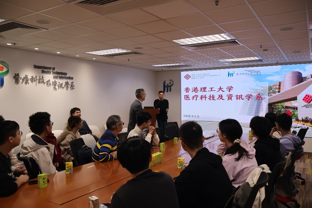
Dr LEUNG Wan Shun introducing the background and history of PolyU and the HTI department
The two parties exchanged gifts and took group photos to commemorate the visit. Representatives from both sides engaged in in-depth discussions, expressing hopes for further strengthening cooperation and the continued success of such exchange activities.
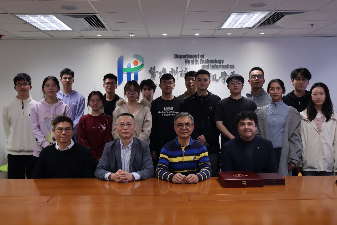
Group photo of faculty and student representatives
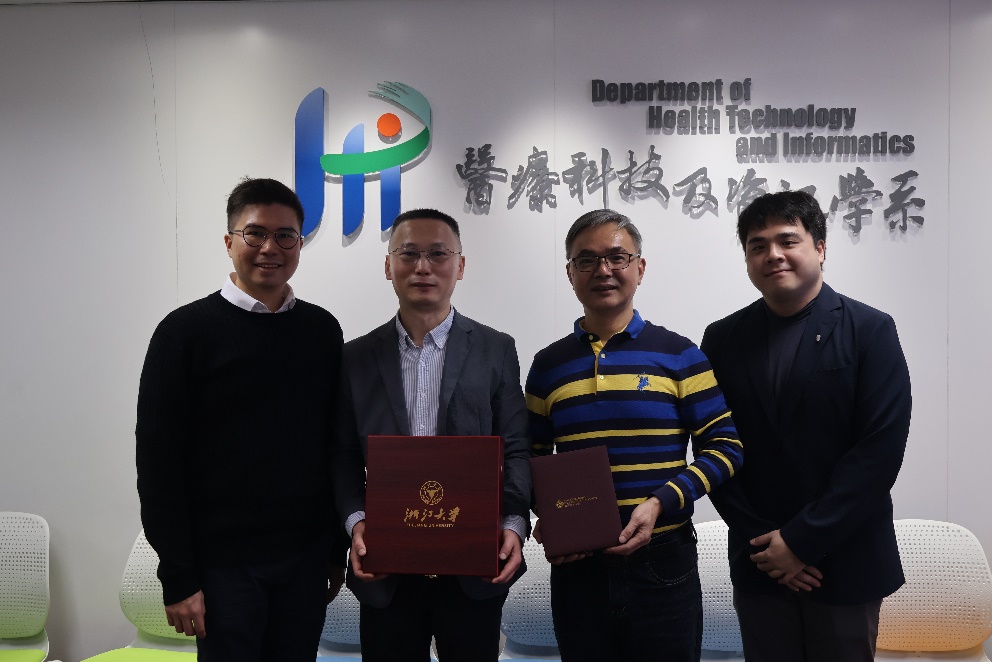
Faculty representatives exchanging college gifts
【New Experiences in Teaching and Experiments】
Dr. Raymond K. H. Hui, Senior Scientific Officer of the teaching laboratories, led the group on a tour of the HTI teaching laboratories. The department boasts numerous well-equipped biological teaching labs, each capable of accommodating 60 students simultaneously for experiments. Remote observation and guidance can also be conducted between labs. In the histology lab, Dr. Raymond explained the structure and use of microtomes and cryostats. Under his guidance, students gained hands-on experience using a cryostat for sectioning. In the cell observation lab, microscopes are connected to a central host system, allowing teachers to check each student's imaging results from the podium and project them for the class. Additionally, Dr. Raymond demonstrated medical testing instruments and clean benches in the teaching labs.

Dr. Raymond guiding a student in using a cryostat
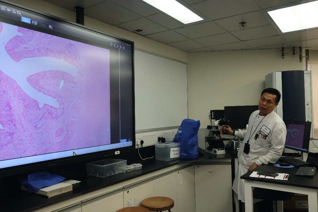
Cell Observation Teaching Laboratory
【New Frontiers in Biomedical Applications】
Students also visited various biomedical research laboratories in groups under the guidance of different professors.
In the pathogen microbiology research group led by Professor SIU Kit Hang, students learned about the close relationship between humans, animals, and the environment, as well as the importance of pathogen and gene detection in animals and the environment for infectious disease surveillance. On the same day, Xia Han, CEO of Yuguo Biotech, visited PolyU HTI to discuss cooperation, and the department organized a seminar. During the seminar, Xia Han introduced the technical capabilities and market share of Yuguo Biotech and their self-developed TB-EASY nucleic acid detection kit for the Mycobacterium tuberculosis complex, expressing a vision to further reduce the number of pulmonary tuberculosis patients per 100,000 people. Students participating in the pathogen microbiology exchange were also invited to attend the seminar.
In the cardiovascular system and metabolism research group supervised by Assistant Professor Cai Yin, students learned the principles, workflows, and application scenarios of Western blotting, as well as important experimental precautions. Under the supervision of doctoral students, they personally tried the gel preparation process. They also observed fluorescence microscopy experiments and understood through practice that different staining methods are required to observe different parts of a cell.
In the hematology research group led by Professor Chien-Ling Huang, students learned about cutting-edge achievements in mouse genetic analysis and observed blood collection and testing techniques firsthand, including the process of spreading blood on slides. Some students also experienced blood analysis operations such as blood drawing, centrifugation, and gel electrophoresis, and observed cell cultures and blood smears under microscopes.
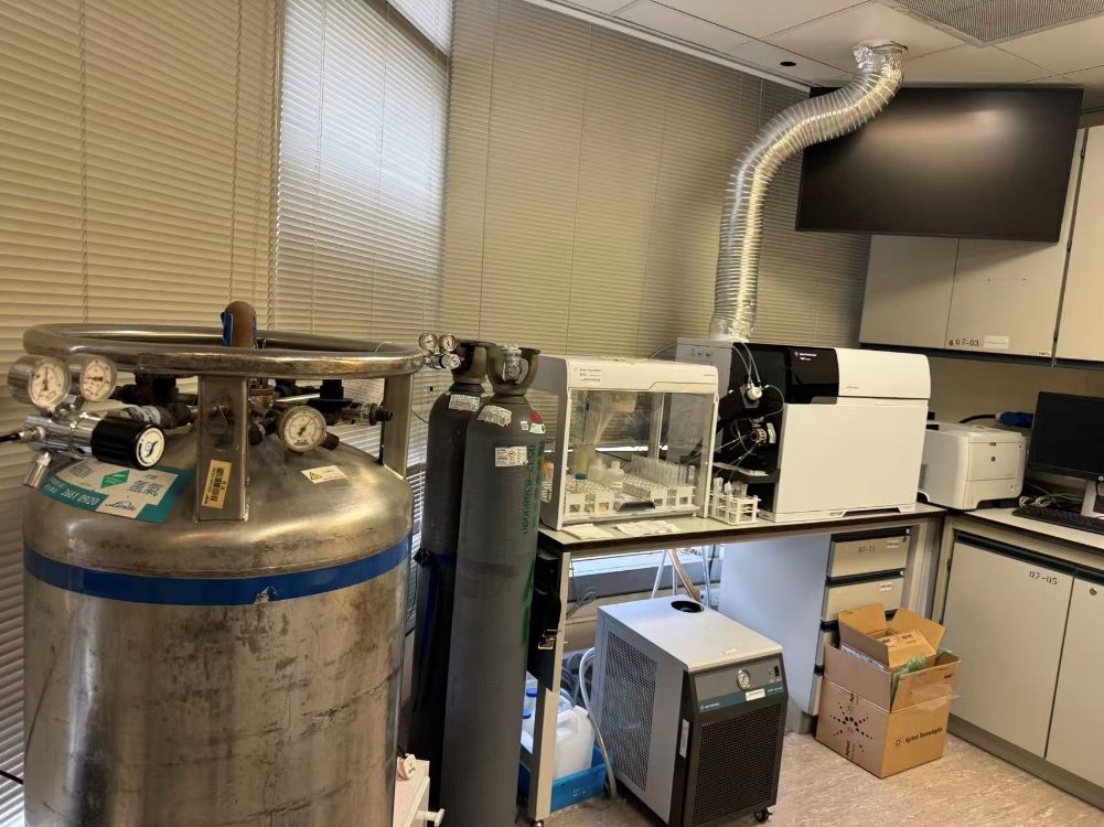
Biomedical research laboratory equipment
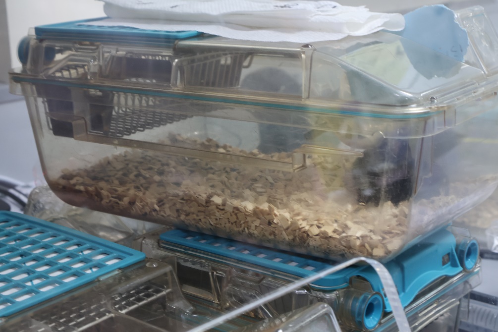
Mouse rearing in the biomedical research laboratory
【New Insights into Radiology and Neuroscience】
Led by Associate Professor of Practice Edward Wong Ting Hei, Edward, the group visited the radiology teaching and research labs to learn about the clinical applications and principles of X-ray and CT equipment. Mr. Wong introduced the core components, mechanisms, and operating procedures of X-ray machines and demonstrated X-ray imaging effects using a mobile phone. He then explained key clinical points, such as adjusting equipment angles for different body parts. Finally, he demonstrated the CT equipment workflow and its imaging capabilities using simulation specimens, emphasizing radiation protection standards and safety protocols.
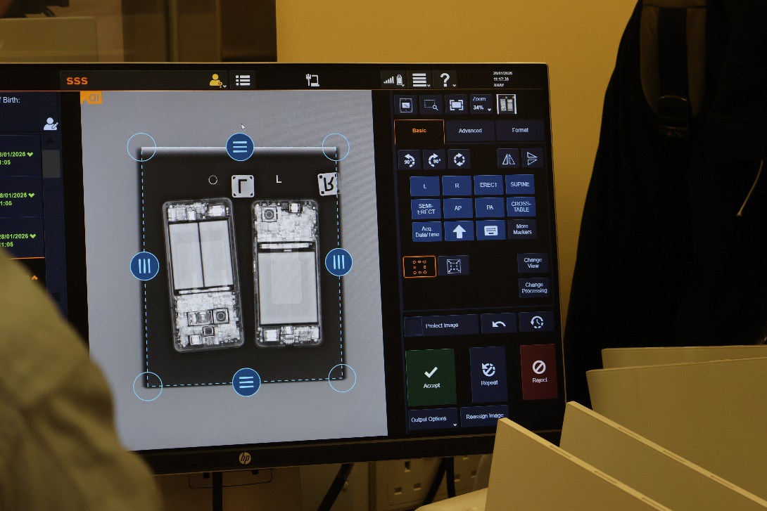
X-ray imaging of mobile phones
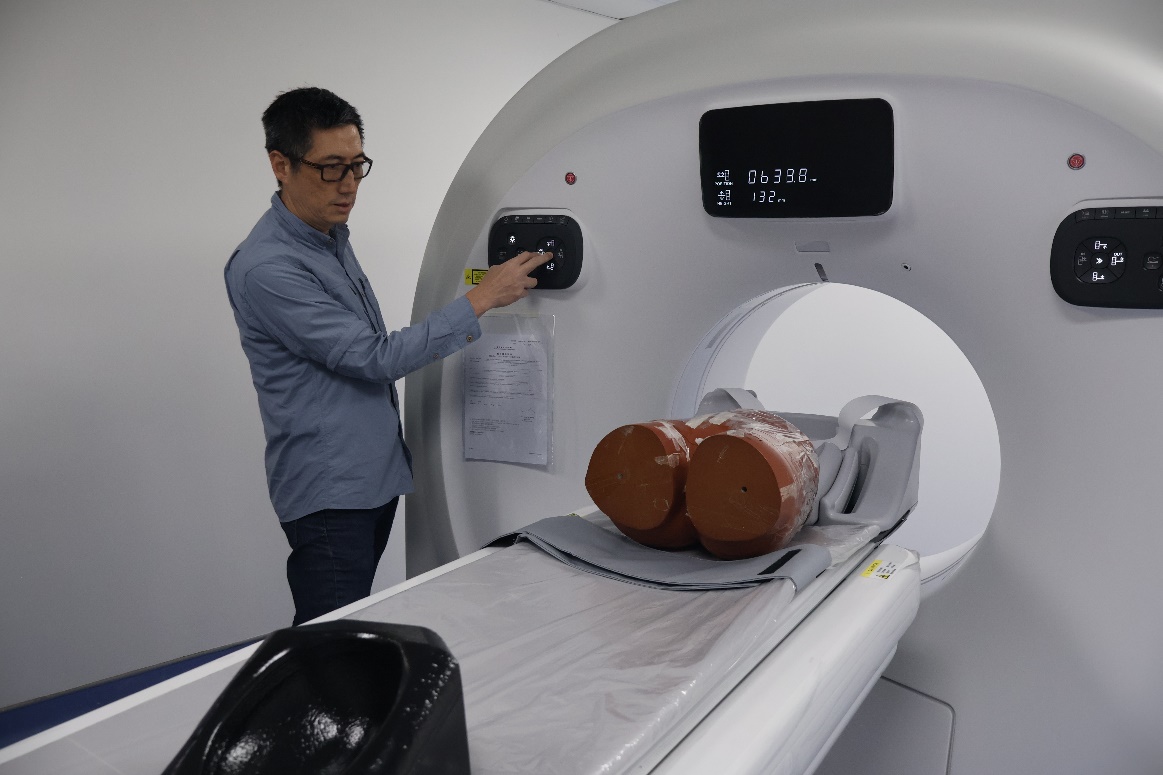
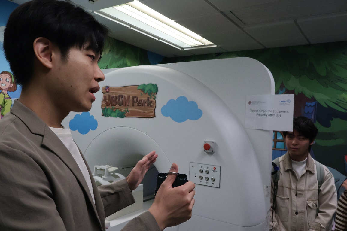
CT simulation specimen demonstration
At the Neuroscience Center Laboratory, students learned neuroscience experimental techniques and equipment applications under the guidance of Dr. Patrick Yeung and other lab members. The lab detailed the structure and positioning principles of the brain stereotaxic instrument. Students then performed hands-on practice on simulation specimens, mastering key steps like skull positioning and coordinate calibration. The lab also features a simulated MRI device to help children adapt to MRI noise. Finally, lab members systematically explained EEG, TMS (Transcranial Magnetic Stimulation), and tDCS (Transcranial Direct Current Stimulation). Student representatives participated in EEG measurements, and lab members provided in-depth explanations of EEG principles and signal processing, effectively bridging classroom knowledge with cutting-edge expansions.
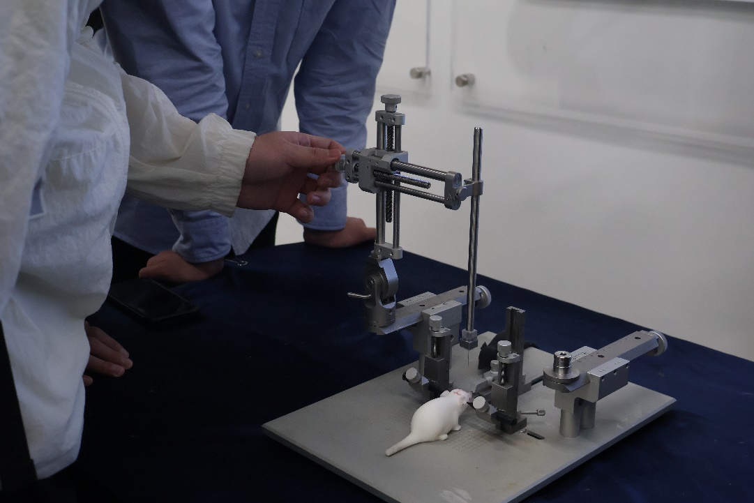
Brain stereotaxic instrument
Simulated MRI equipment
EEG Laboratory
In the radiology lab, Associate Professor of Practice Dr LEUNG Wan Shun introduced radiotherapy and its applications. Professor Leung explained the global use of radiotherapy and the relative shortage of related equipment and personnel. He used various simulation devices to show how radiation attenuates in human tissue and posed questions to stimulate analytical thinking among the students.
Radiology simulation demonstration
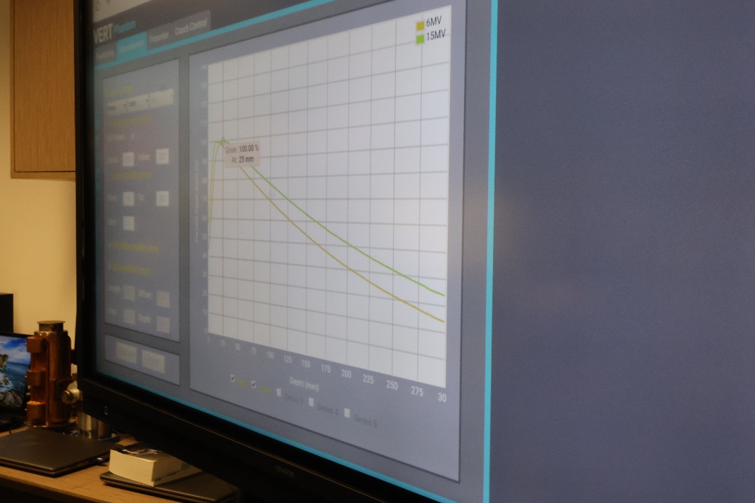
Radiology numerical simulation
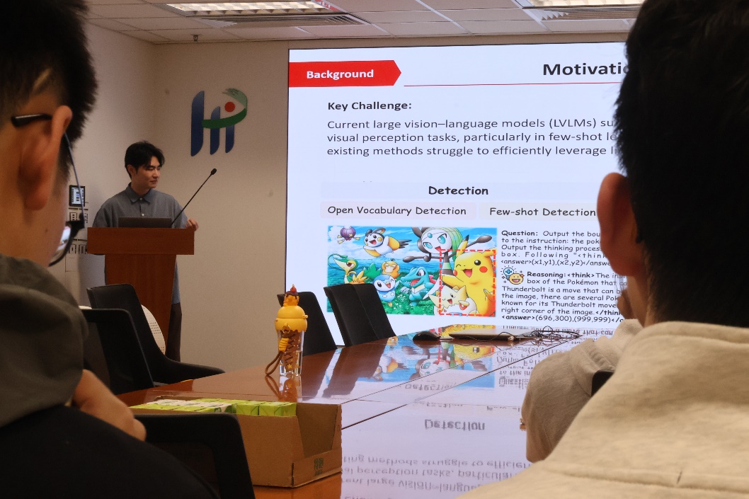
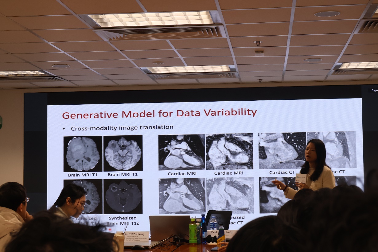
【New Depths in Literature Discussion and AI】
Students participated in a Journal Club organized by Professor Qiu Anqi's research group. They exchanged ideas on cutting-edge literature involving the intersection of Artificial Intelligence (such as Large Language Models and Computer Vision) and Biomedical Engineering. Following the discussion, Professor Qiu shared insights on future studies, career choices, and research planning, answering students' questions regarding school selection, time management, and research progress. Students also attended an academic lecture on AI in medical research, covering topics like generative AI systems, integrated AI models for medical imaging and clinical text, and data preprocessing using generative AI or Fourier transform.
Knowledge sharing and exchange with Professor Qiu Anqi's research group
Academic lecture on the frontier of AI-Medical intersection